Home
/ I. Identify The Main Muscles Of The Body, Using The Accompanying Diagram; Indicate, Using The Letters Provided, Where Each Muscle Group Is On The Diagram. ~ Skeletal System Building A Medical Terminology Foundation
I. Identify The Main Muscles Of The Body, Using The Accompanying Diagram; Indicate, Using The Letters Provided, Where Each Muscle Group Is On The Diagram. ~ Skeletal System Building A Medical Terminology Foundation
I. Identify The Main Muscles Of The Body, Using The Accompanying Diagram; Indicate, Using The Letters Provided, Where Each Muscle Group Is On The Diagram. ~ Skeletal System Building A Medical Terminology Foundation. Start studying muscles of the human body (anterior). A basic human skeleton is studied in schools with a simple diagram. The substance labeled catalyst is also known as a)molecular size b)physical shape c)carrying capacity d)stored energy 4.the function of a specific enzyme is most directly influenced by its a)different kinds of body cells in a cloned sheep Find the indicated structures in the diagrams provided, based on the directional terms given. Superficial and deep anterior muscles of upper body
Cranial aponeurosis buccinator frontalis masseter platysma occipitalis orbicularis oculi orbicularis oris. Muscles, the specialized tissues that facilitate body movement, make up about 40% of body weight. Enter the correct letter in the answer blank. Skin diagram, chapter 12 11 terms. Muscles of the arm and forearm 24.
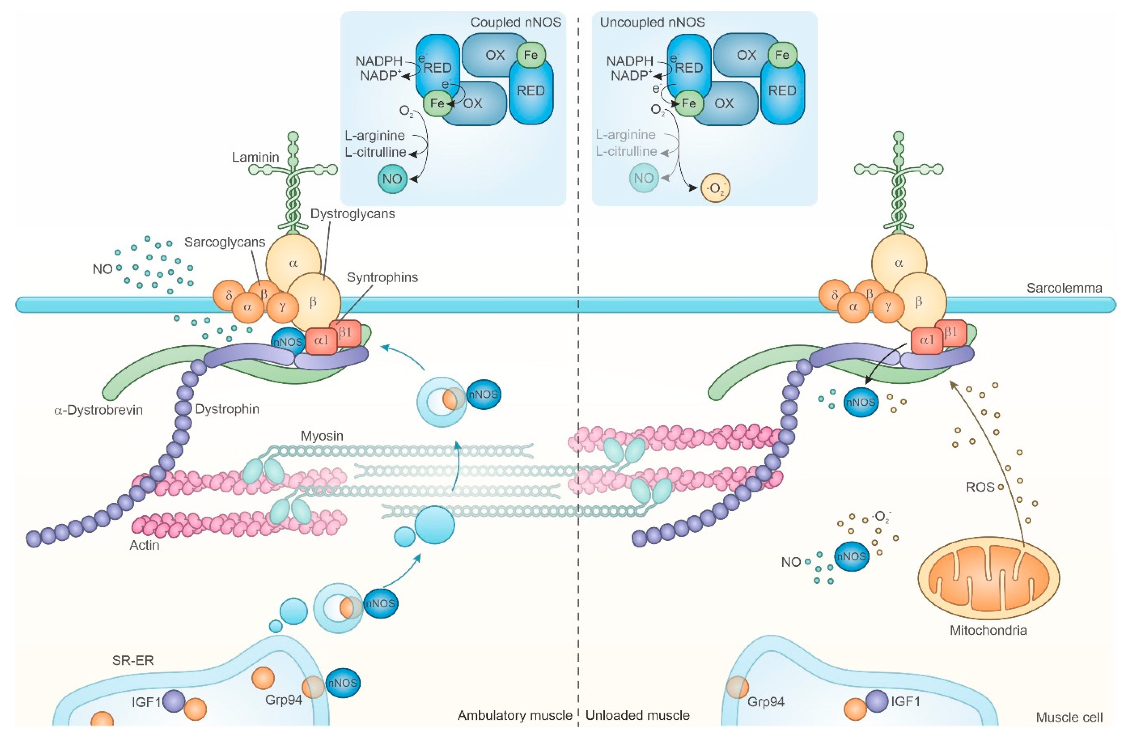
Cells Free Full Text Master Regulators Of Muscle Atrophy Role Of Costamere Components Html from www.mdpi.com Meniscus in a knee joint 2. The compartments of the distal upper extremity include the superficial anterior, deep anterior, and posterior regions. Indicate, using the letters provided, where each muscle group is on the diagram. 3.the diagram below illustrates a biochemical process that occurs in organisms. Major surface muscles in the body surface muscles are the muscles that you can see on the superficial layer of the body. Learn vocabulary, terms, and more with flashcards, games, and other study tools. Integumentary system, integumentary, integumentary, integumentary. Enter the correct letter in the answer blank.
Pulmonary circuit and systemic circuit.
A keratinocyte is a cell that manufactures and stores the protein keratin. Cranial aponeurosis buccinator frontalis masseter platysma occipitalis orbicularis oculi orbicularis oris. Forms the walls of the 8. The cells in all of the layers except the stratum basale are called keratinocytes. Dorsal refers to the _____ of the human body, while ventral refers to the _____ of the human body. Indicate, using the letters provided, where each muscle group is on the diagram. Epithelial tissue is made of layers of cells that cover the surfaces of the body that come into contact with the exterior world, line internal cavities, and form. A muscle group consisting of four muscles that is located along the front of the thigh. As shown in figure 9.1, all of these connective tissue sheaths are continuous with one another as well as with the tendons that join muscles to bones. This is opposed to other components or tissues in muscle such as tendons or perimysium. Meniscus in a knee joint 2. The structure to find will be one of those at the end of an unlabeled line. Learn vocabulary, terms, and more with flashcards, games, and other study tools.
There are additional body cavities which we will only discuss in lecture. Connects the ribs to the sternum 3. Click on the name of the muscle, or the image, to see weight training exercises. The pump for the pulmonary circuit, which circulates blood through the lungs, is the right ventricle.the left ventricle is the pump for the systemic circuit, which provides the blood supply for the tissue cells of the body. This is opposed to other components or tissues in muscle such as tendons or perimysium.
Skeletal System Building A Medical Terminology Foundation from ecampusontario.pressbooks.pub The other end of the muscle stays fixed and the part of the muscle that moves is moved towards this fixed point. Amino acids are organic molecules that, when linked together with other amino acids, form a protein.amino acids are essential to life because the proteins they form are involved in virtually all cell functions. The compartments of the distal upper extremity include the superficial anterior, deep anterior, and posterior regions. Microscopic anatomy of skeletal muscle 4. Find the indicated structures in the diagrams provided, based on the directional terms given. Muscles of the arm and forearm 24. The structure to find will be one of those at the end of an unlabeled line. Enter the correct letters (or terms if desired) in the answer blanks.
This type of muscle creates movement in the body.
When muscle fibers contract, they pull on these sheaths, which transmit the pulling force to the bone to be moved. Using the terms provided above, identify the muscles described next. This article explains the bone structure of the human body, using a labeled skeletal system diagram and a simple technique to memorize the names of all the bones. Label the extensor digitorum muscle in the figure below. Meniscus in a knee joint 2. Start studying muscles of the human body (anterior). Muscles of the arm and forearm 24. There are additional body cavities which we will only discuss in lecture. The structure to find will be one of those at the end of an unlabeled line. Skin diagram, chapter 12 11 terms. Amino acids are organic molecules that, when linked together with other amino acids, form a protein.amino acids are essential to life because the proteins they form are involved in virtually all cell functions. Dorsal refers to the _____ of the human body, while ventral refers to the _____ of the human body. Using choices from the list at the right, correctly identify the muscles provided with leader lines on the diagram.
Integumentary system, integumentary, integumentary, integumentary. Muscles are generally attached at two points in the body. Enter the correct letters (or terms if desired) in the answer blanks. Using choices from the list at the right, correctly identify the muscles provided with leader lines on the diagram. It is important to know the origin, insertion, and actions of some of these major muscles in the body.
Names Functions And Locations Of Cranial Nerves from www.thoughtco.com Start studying muscles of the human body (anterior). All the major muscle groups of the body from front and back. Epithelial tissue is made of layers of cells that cover the surfaces of the body that come into contact with the exterior world, line internal cavities, and form. The cells in all of the layers except the stratum basale are called keratinocytes. The blood vessels of the body are functionally divided into two distinctive circuits: The pump for the pulmonary circuit, which circulates blood through the lungs, is the right ventricle.the left ventricle is the pump for the systemic circuit, which provides the blood supply for the tissue cells of the body. The four types of tissues in the body are epithelial, connective, muscle, and nervous. Cranial aponeurosis buccinator frontalis masseter platysma occipitalis orbicularis oculi orbicularis oris.
Dorsal refers to the _____ of the human body, while ventral refers to the _____ of the human body.
Epithelial tissue is made of layers of cells that cover the surfaces of the body that come into contact with the exterior world, line internal cavities, and form. This diagram depicts muscle of the body diagrams 744×1054 with parts and labels. When muscle fibers contract, they pull on these sheaths, which transmit the pulling force to the bone to be moved. This type of muscle creates movement in the body. Muscle group that extends the thigh and flexes the knee. There are additional body cavities which we will only discuss in lecture. A keratinocyte is a cell that manufactures and stores the protein keratin. Identify the four types of tissue in the body, and describe the major functions of each tissue. Muscles, the specialized tissues that facilitate body movement, make up about 40% of body weight. Other sets by this creator. One end is pulled by the muscle to create movement. The diagram below summarizes the transfer of energy that eventually powers muscle activity. It is important to know the origin, insertion, and actions of some of these major muscles in the body.
Share :
Post a Comment
for "I. Identify The Main Muscles Of The Body, Using The Accompanying Diagram; Indicate, Using The Letters Provided, Where Each Muscle Group Is On The Diagram. ~ Skeletal System Building A Medical Terminology Foundation"
/cranial-nerves-2-1e3d489c9104495dbcc609ea188af32d.jpg)

Post a Comment for "I. Identify The Main Muscles Of The Body, Using The Accompanying Diagram; Indicate, Using The Letters Provided, Where Each Muscle Group Is On The Diagram. ~ Skeletal System Building A Medical Terminology Foundation"